Overview
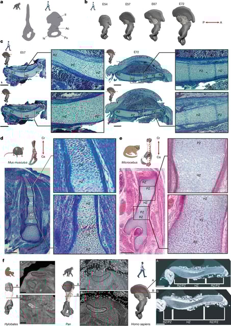
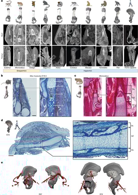
- A Nature study details a 90‑degree reorientation of ilium cartilage around gestational day 53 followed by a delayed, rearward ossification that locks in a short, wide pelvis.
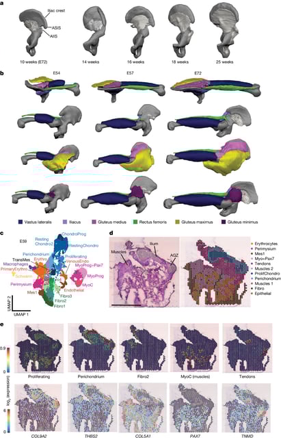
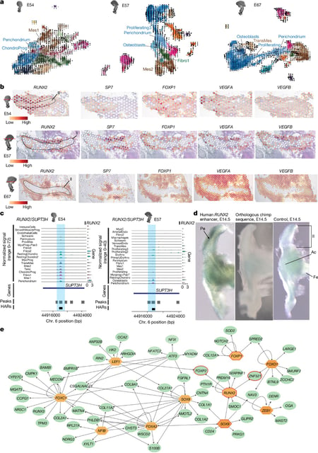
- Single‑cell and spatial transcriptomics reveal a network of 300‑plus regulators, with SOX9 and PTH1R implicated in the growth‑plate flip and RUNX2 in the ossification delay.
- Researchers examined 128 human embryonic samples and embryos from nearly two dozen primate species using histology, CT imaging, and museum plus clinical tissue collections.
- The team proposes the growth‑axis change arose near the human–chimp split 5–8 million years ago, with the ossification shift emerging within the past ~2 million years, aligning with Ardipithecus and Australopithecus pelves.
- Authors say these steps enabled efficient, habitual bipedalism and a birth canal suited to big‑brained infants, informing updated models of locomotion and childbirth evolution.