Overview
- Published on July 31, 2025 in Analytical Chemistry, this work is the first demonstration of concurrent cryo-EM structural imaging and elemental mapping in organic nanomaterials.
- By integrating the three-window method and drift compensation, the approach minimizes ice-plasmon background signals and sample blur during electron energy loss spectroscopy.
- A newly released ParallEM control system extension automates energy-shift adjustments, reducing manual intervention and enhancing reproducibility.
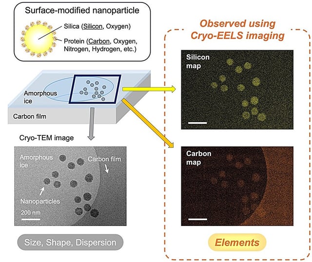
- Validation on silica and hydroxyapatite nanoparticles showed clear visualization of silicon, calcium and phosphorus distributions at roughly 10 nm resolution.
- This low-dose elemental mapping technique is poised to accelerate research across biomaterials, medical implants, catalysts, food science and functional inks.