
Overview
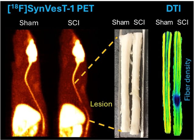
- In a T7 contusion model, [18F]SynVesT-1 uptake at the injury epicenter fell about 61% on day 1 and 53% on days 9–11 versus sham controls.
- PET also detected reduced SV2A signal in remote brain regions, including the amygdala, limbic insular cortex, and cerebellum.
- Ex vivo diffusion tensor imaging identified fiber damage in the internal capsule and somatosensory cortex, aligning with the PET results.
- Molecular assays, including immunohistochemistry and Western blotting, confirmed SV2A loss at the lesion site.
- The authors propose SV2A PET as a noninvasive quantitative biomarker for monitoring spinal cord injury and assessing treatments, with disclosures noting related patents and company affiliations.