Overview
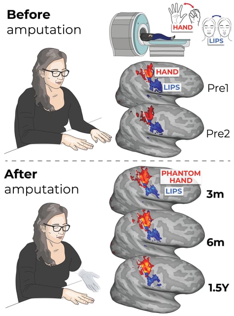
- Published in Nature Neuroscience, a longitudinal fMRI study of three patients scanned before surgery and up to five years after hand removal found stable cortical representations of the hand and lips.
- A machine-learning decoder trained on pre-amputation finger patterns correctly identified which phantom finger participants attempted to move post-surgery, indicating preserved finger-specific codes.
- Imaging showed no takeover of the missing hand’s cortical area by neighboring regions such as the lips, contradicting long-held remapping theories.
- Comparisons with 26 long-term upper-limb amputees revealed similar stability of hand and lip representations decades after limb loss.
- The findings support long-term viability for neural prosthetics and shift phantom limb pain strategies toward targeting peripheral nerve changes, though experts note the small longitudinal sample and urge larger, more diverse cohorts.