
Overview
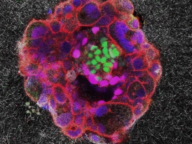
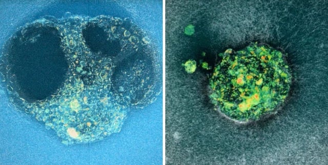
- The peer-reviewed study published in Science Advances presents time-lapse fluorescence recordings capturing human embryo implantation every 20 minutes over 16–24 hours within a collagen-rich 3D gel.
- Human blastocysts actively generate traction forces to invade and carve into the uterine-like matrix, in contrast to mouse embryos that rely on uterine folding to accommodate implantation.
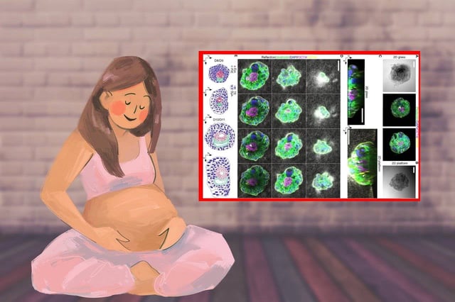
- Measurement of collagen fiber displacement provides a “mechanical footprint” of implantation, offering potential metrics to refine embryo selection and culture conditions in assisted reproduction.
- Developed by IBEC with partners including Hospital Universitario Dexeus, IDIBELL and the University of Tel Aviv, the collagen-based platform mimics uterine tissue outside the maternal body but remains confined to in vitro conditions.
- Clinical translation will require further validation, ethical approvals and clinical research before these mechanobiological insights can improve IVF practices.