Overview
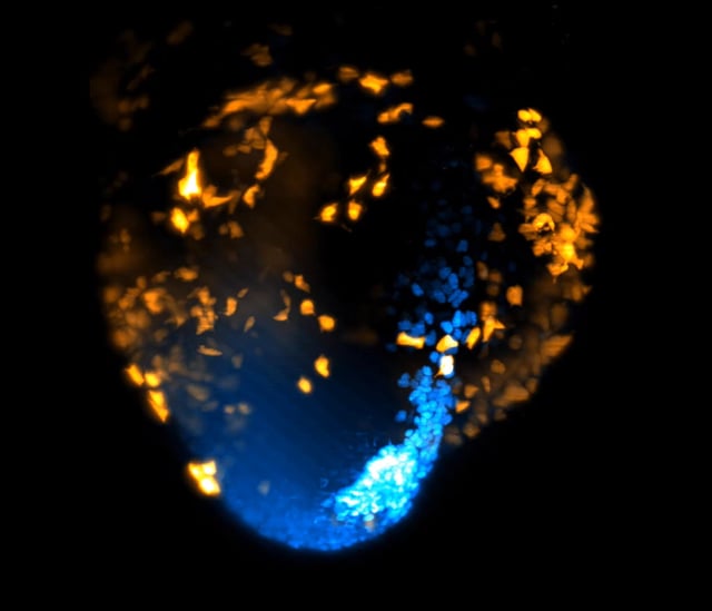
- Researchers at UCL and the Francis Crick Institute recorded the first-ever real-time 3D footage of heart formation in a living mouse embryo.
- The study utilized advanced light-sheet microscopy, a non-invasive imaging technique, to capture detailed time-lapse images over 40 hours without damaging tissue.
- Findings show that cardiac cell fate determination and organized migration begin within hours of gastrulation, challenging long-standing models of heart development.
- The heart forms from distinct cell groups appearing at different times and locations, coordinated by previously unknown signals during early development.
- Published in EMBO Journal and funded by the British Heart Foundation, the research could transform understanding and treatment of congenital heart defects and advance tissue engineering.