Overview
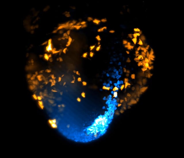
- Using advanced light-sheet microscopy, researchers at UCL and the Francis Crick Institute visualized the earliest stages of heart formation in a live mouse embryo over 40 hours.
- The study revealed that cardiac progenitor cells differentiate and follow precise migration paths much earlier than previously thought, within hours of gastrulation.
- Distinct cell groups were shown to contribute to heart formation, challenging longstanding models of stochastic and unsynchronized early development.
- These findings could revolutionize approaches to treating congenital heart defects, which affect nearly 1% of newborns globally, and advance regenerative medicine efforts.
- The research, supported by the British Heart Foundation, was published in The EMBO Journal and marks a significant step in understanding organogenesis through non-invasive imaging.