Overview
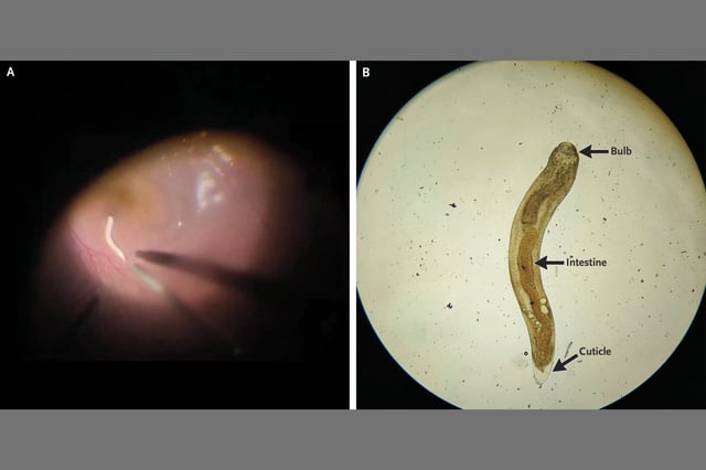
- A 35-year-old man from rural central India endured eight months of redness, blurred vision and panuveitis before funduscopy revealed a sluggish worm in his posterior segment.
- Ophthalmologists performed a pars plana vitrectomy to suction out the live intraocular larva.
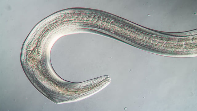
- Microscopic analysis of the extracted specimen confirmed Gnathostoma spinigerum based on its cephalic bulb, thick cuticle and well-defined intestine.
- Oral and ocular glucocorticoids plus albendazole resolved the infection, but at eight weeks a postoperative cataract limited vision in the affected eye to 20/40.
- Researchers urge clinicians in endemic areas to consider ocular gnathostomiasis in patients with persistent uveitis and histories of undercooked freshwater fish or poultry consumption.


