Overview
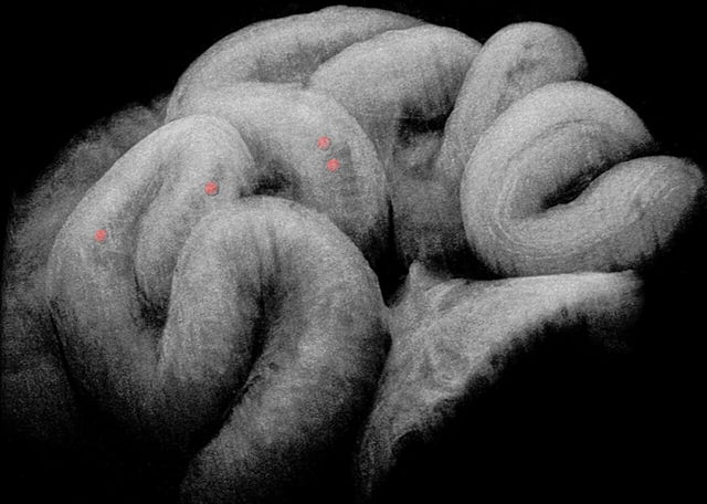
- Researchers at Stevens Institute used an implantable optical window and label-free 3D and 4D OCT to directly visualize mouse oviduct dynamics in vivo.
- High-speed imaging captured coordinated cilia beating and muscular contraction waves traveling from the ampulla through the isthmus.
- Detailed spatiotemporal analysis showed the oviduct functions as a leaky peristaltic pump, pushing fluid forward and creating suction at relaxation sites to move embryos.
- Occasional constrictions at oviduct bend points halted backward movement, resulting in slow net displacement of embryos toward the uterus.
- Building on these findings, the team is now planning in vivo studies to investigate abnormal transport mechanisms underlying tubal ectopic pregnancy and infertility.