Particle.news

Download on the App Store

New Two-Photon Microscope Revolutionizes Real-Time Neural Imaging

Innovative adaptive sampling and line illumination techniques offer unprecedented insights into brain function and neurological diseases.

Image
A new two-photon fluorescence microscope can capture high-speed images of neural activity at cellular resolution thanks to a new adaptive sampling scheme and line illumination. The illustration shows the adaptive sampling scheme, in which a laser beam patterned by a digital micromirror device selectively illuminates neurons in the brain tissue to image their activity. Credit: Wei Wei and Mei Xueting, LINGO.AI LLC, edited

Overview

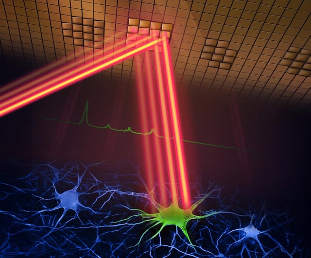
  • The microscope captures high-speed images of neural activity at cellular resolution, significantly faster than traditional methods.
  • Adaptive sampling and line illumination reduce laser power, minimizing damage to brain tissue.
  • Researchers can now observe real-time neural dynamics, crucial for understanding learning, memory, and decision-making.
  • The technology holds potential for early-stage study and treatment of neurological diseases like Alzheimer's and Parkinson's.
  • Future enhancements aim to integrate voltage imaging and improve the microscope's usability and size.