Overview
- Okayama University and Maastricht University researchers report a sample-preparation and imaging method in Scientific Reports that correlates lipid identity with precise anatomical location in intact worms.
- The workflow aligns nematodes on a custom microfluidic chip, embeds them in gelatin–carboxymethyl cellulose, cryosections them, and applies MALDI-MSI alongside Oil Red O staining.
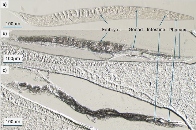
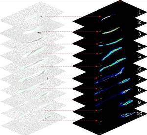
- Stacked sectional data produce three-dimensional reconstructions showing region-specific lipid patterns across the pharynx, intestine, reproductive system, and embryos.
- A lipid linked to cholesterol metabolism localized mainly to the pharynx and anterior intestine, highlighting tissue-specific roles in nutrient handling.
- Technical reproducibility was high, with biological differences between individual worms exceeding methodological variability, and the team plans to extend the method to disease-related strains and add quantification tools.