Overview
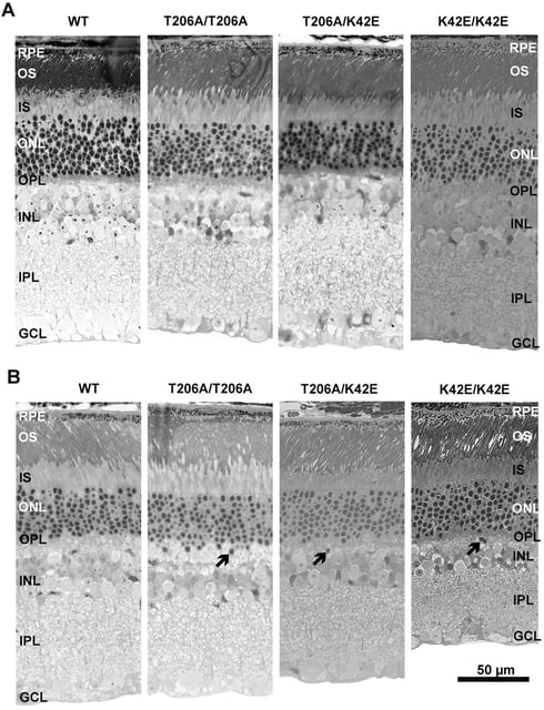
- Researchers at UAB report T206A/K42E and T206A/T206A Dhdds knock-in mice that at 12 months display retinal changes comparable to the established K42E/K42E model.
- Observed changes include inner nuclear layer thinning, reduced bipolar and amacrine cell densities, and electroretinography b‑wave reduction with preserved a‑waves.
- The T206A/T206A genotype has not been identified in humans, yet its similar phenotype indicates the T206A allele is pathogenic in mice.
- The team highlights T206A/K42E as a common patient variant, making these models pertinent for studying RP59 mechanisms.
- The findings are published in Disease Models & Mechanisms with authors from UAB, the University of Oklahoma Health Sciences Center, and the VA Western New York Healthcare System.

