Overview
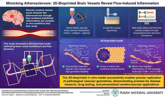
- Researchers from Pusan National University and POSTECH report a validated in vitro model of narrowed brain vessels published in Advanced Functional Materials on June 24, 2025.
- They used embedded coaxial bioprinting and a hybrid bioink of porcine aorta dECM, collagen, and alginate to fabricate perfusable conduits with controlled luminal narrowing.
- Human endothelial cells, including HUVECs and HBMECs, formed continuous linings that expressed CD31, VE‑cadherin, and ZO‑1 and exhibited selective permeability indicating barrier integrity.
- Computational fluid dynamics and tracer bead tests confirmed stenosis‑induced disturbed flow, and the model showed significant upregulation of inflammatory markers under these conditions.
- The team positions the platform for drug screening, toxicity testing, and personalized models, with planned enhancements using brain‑specific ECM, vascular support cells, patient‑derived tissues, and organ‑on‑chip integration.