Overview
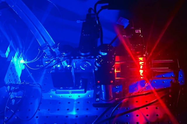
- MIT researchers developed a streamlined technique combining a suspended microchannel resonator (SMR) with fluorescence microscopy to measure single-cell density at unprecedented speeds.
- The system achieves throughput of up to 30,000 cells per hour, overcoming previous limitations of time-consuming dual-fluid measurements.
- Density changes were validated as biomarkers for cellular states, with studies showing T cell activation decreases density due to increased water content.
- Preliminary results from Travera indicate that combining mass and density metrics improves predictions of T cell responses to immunotherapy drugs.
- The technique also distinguishes pancreatic cancer cells' susceptibility to chemotherapy within days, offering potential for rapid precision oncology assessments.
