Overview
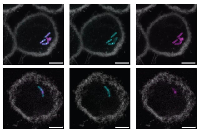
- MIT chemists have created an oxaziridine-based probe that targets the ManLAM glycan in Mycobacterium tuberculosis, enabling real-time visualization of its cell wall distribution.
- The probe demonstrates high specificity by selectively labeling the sulfur-containing MTX sugar in ManLAM, with no fluorescent signal detected in nonpathogenic Mycobacterium smegmatis.
- The study, published in the Proceedings of the National Academy of Sciences, reveals that ManLAM remains attached to the bacterial cell wall during early macrophage infection, challenging previous hypotheses of glycan shedding.
- Researchers aim to translate this breakthrough into rapid, urine-based TB diagnostics that could provide a faster and more accessible alternative to sputum culture and imaging methods.
- This innovation could also be used to monitor antibiotic efficacy and immune responses, offering new insights into bacterial pathogenesis and treatment strategies.
