Overview
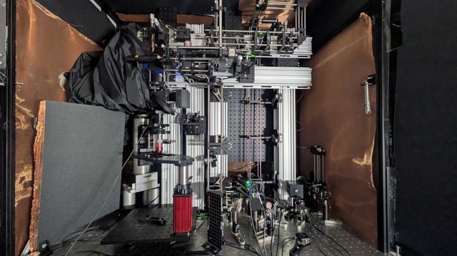
- The ‘Multiphoton-In and Acoustic-Out’ system uses three-photon excitation to induce localized thermal expansion in cells and acoustic detection to avoid external labels.
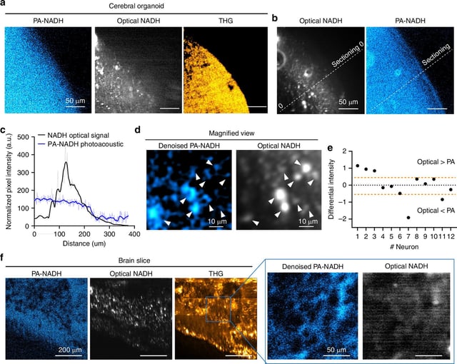
- Researchers detected NAD(P)H molecules through 1.1 mm-thick cerebral organoids and 0.7 mm mouse brain slices, exceeding previous depth limits by more than fivefold.
- A sensitive ultrasound transducer captures the sound waves generated by molecular excitation and software converts them into high-resolution images.
- To enable in vivo imaging, engineers are redesigning the detector placement so that both light source and microphone sit on the same side of living tissue.
- The label-free approach could monitor biomarkers such as NAD(P)H and GCaMP in real time during neuroscience experiments or human brain surgeries.