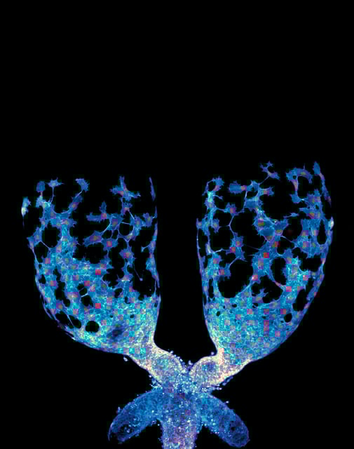
Overview
- UNC researchers used advanced live imaging to show muscle precursor mesenchymal cells crawling collectively over the developing fruit fly testis, sculpting its transformation from oval to spiral.
- The Plexin/Semaphorin signaling pathway regulates the balance between epithelial and mesenchymal states and is essential for coordinated cell migration during organ molding.
- This dynamic behavior challenges the view of mesenchymal cells as passive supporters by demonstrating their active architectural role in organogenesis.
- Findings suggest that the molecular machinery guiding normal tissue sculpting may mirror mechanisms of cancer cell invasion and metastasis.
- Employing Drosophila melanogaster allowed high-resolution visualization and genetic manipulation, underscoring the model’s relevance to human developmental biology.