
Overview
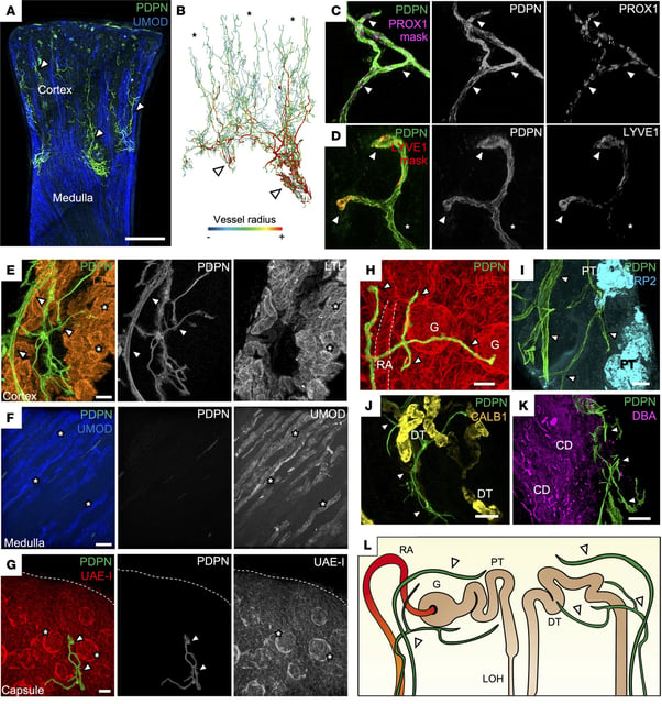
- Teams at the Wellcome Sanger Institute, UCL and the University of Cambridge combined single-cell RNA sequencing with advanced 3D imaging to map kidney lymphatics in healthy and rejected human tissue.
- During rejection, lymphatic vessels expand into the kidney medulla—which normally lacks these vessels—and become structurally disorganized.
- Endothelial junctions on lymphatic vessels shift from looser “button” formations to tighter “zipper” configurations associated with reduced immune cell escape.
- Immune balance around the vessels is disrupted, as T cells trigger braking signals while harmful antibodies and other immune cells attack the graft and show evidence of targeting the vessels themselves.
- Published September 16, 2025 in the Journal of Clinical Investigation, the findings reposition lymphatics as contributors to chronic rejection and highlight preclinical targets, against persistent long-term graft failure and a UK waiting list exceeding 8,000 people.