Overview
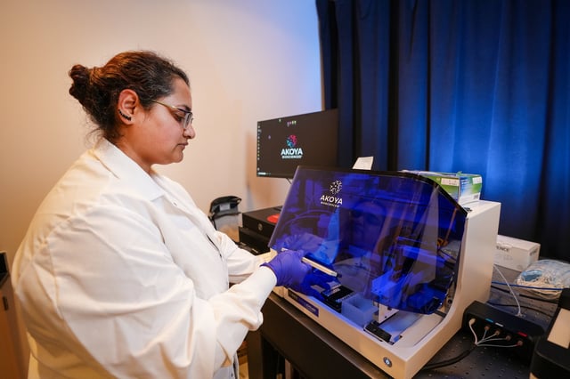
- Indiana University scientists have pioneered a non-disruptive imaging method to study intact mouse bone marrow using Phenocycler 2.0, visualizing 25 cellular markers simultaneously.
- The technique preserves tissue integrity and spatial context, enabling deeper insights into bone marrow's cellular architecture and its role in disease mechanisms.
- This is the first successful application of Phenocycler 2.0 to bone marrow imaging, overcoming limitations of traditional methods like flow cytometry and standard fluorescence microscopy.
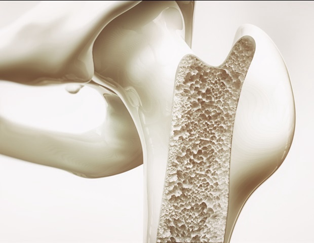
- The innovation could accelerate drug discovery and improve therapies for conditions such as blood cancers, autoimmune diseases, and musculoskeletal disorders.
- The methodology has been published in the journal Leukemia, with a provisional patent filed as researchers work to expand the marker panel for broader preclinical applications.