Overview
- CSHL graduate students Steven Lewis, Lucia Téllez Pérez, and Samantha Henry developed the system in the dos Santos lab after Lewis drew on plant branching models presented by Saket Navlakha.
- The method is published in the Journal of Mammary Gland Biology and Neoplasia with DOI 10.1007/s10911-025-09589-1.
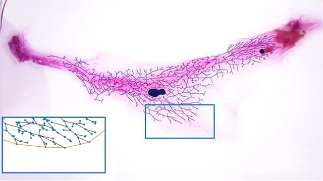
- Users trace duct structures from stained images and employ NetworkX to render node-and-edge graphs that enable automated measurements of total length, ducts, alveoli, and branch points.
- The pipeline targets a faster, more consistent alternative to labor-intensive tissue slicing and manual counting that can miss whole-gland architecture.
- Validation is limited to mice for now, and the team says the code could be adapted to other branching systems and used in research on risk factors and potential early warning signals.