Overview
- Scientists implanted a glass-resin window in live mice and applied label-free 3D and 4D optical coherence tomography to capture oviduct biomechanics and preimplantation embryo movement in real time.
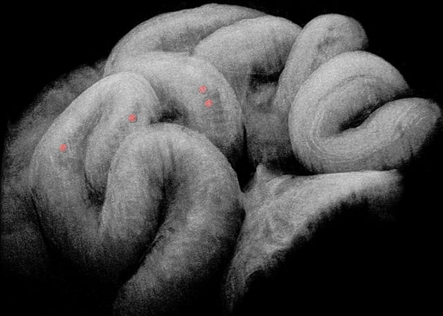
- Quantitative spatiotemporal analysis revealed the fallopian tube functions as a leaky peristaltic pump that pushes fluid forward with contraction waves and pulls it back through relaxation to achieve net embryo displacement toward the uterus.
- Muscular contraction waves were observed originating in the ampulla and propagating through the isthmus while ciliary beat frequency was inferred from OCT intensity fluctuations to complement muscle-driven transport.
- This study marks the first in vivo use of advanced OCT to directly observe preimplantation embryo transport mechanics in the mammalian oviduct.
- Building on these findings, the team is now initiating follow-up imaging studies to probe abnormal transport dynamics that can cause tubal ectopic pregnancy and related fertility disorders.