Overview
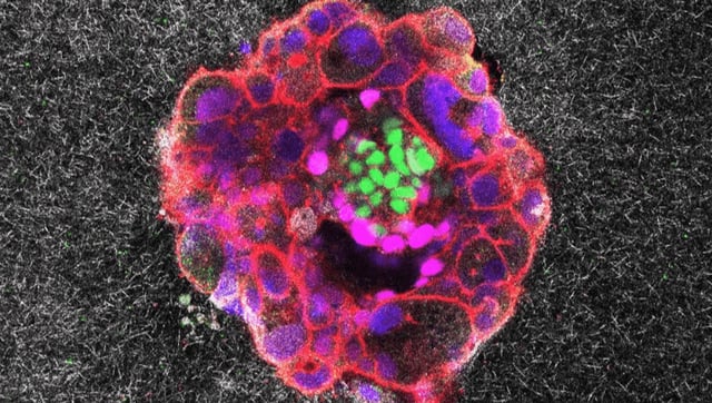
- The IBEC team engineered 2D and 3D collagen-protein matrices that simulate uterine outer layers to enable time-lapse observation of blastocyst implantation.
- In the 2D setup 72% of human blastocysts attached and invaded up to 200 µm into the gel, while the 3D configuration achieved an 80% survival and invasion rate.
- Traction force microscopy captured pulsatile pulling forces exerted by embryos, showing that these mechanical cues continuously remodel the surrounding matrix during invasion.
- Comparative tests demonstrated that mouse blastocysts remained surface-limited, whereas human embryos fully burrowed into and integrated with the simulated uterine tissue.
- Researchers are now pursuing theoretical modelling and follow-up experiments to probe initial attachment and add maternal components, highlighting both IVF promise and model limitations alongside disclosed commercial partnerships.